Nikon Announces the Winners of its “Small World” Competition
See a selection of beautiful images captured by scientists gazing through light microscopes
![]()
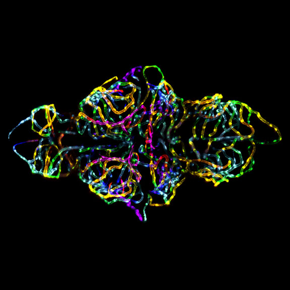
1st Place: The blood-brain barrier in a live zebrafish embryo. Image by Dr. Jennifer L. Peters and Dr. Michael R. Taylor.
Last week, Nikon unveiled the winners of its 38th annual Small World Photomicrography Competition. What’s photomicrography, you ask? Well, while there are many techniques involved, the genre, simply put, is photography captured through a light microscope.
Researchers use photomicrographs as a means of scientific inquiry. The images depict life in all its glorious, magnified detail. “But a good photomicrograph is also an image whose structure, color, composition and content is an object of beauty, open to several levels of comprehension and appreciation,” reads the competition’s Web site.
For its 2012 contest, Nikon received more than 2,000 submissions—stunning images of algae, insects, seeds, snowflakes, embryos and minerals—from photomicrographers around the world. Judges plucked from the cell biology departments at Northwestern University and Columbia University and the staffs of Popular Science and the scientific journal Nature Methods then selected 115 finalists “on the basis of originality, informational content, technical proficiency and visual impact,” according to the site. Those finalists were further divided into 20 top winners, 11 honorable mentions and 84 images of distinction.
First-place winners Jennifer Peters and Michael Taylor, both of St. Jude Children’s Research Hospital in Memphis, Tennessee, achieved a photomicrography first. Their winning entry, “the blood-brain barrier in a live zebrafish embryo,” pictured above, is believed to be the first image to show the creation of this barrier, between circulating blood and fluids in the central nervous system, in a living organism.
“We used fluorescent proteins to look at brain endothelial cells and watched the blood-brain barrier develop in real-time,” said Peters and Taylor in a press release. “We took a three-dimensional snapshot under a confocal microscope. Then, we stacked the images and compressed them into one—pseudo coloring them in rainbow to illustrate depth.”
Nikon launched a Popular Vote contest on Facebook, to determine a fan favorite. Which of the finalists do you like best? Polls are open until November 13, and the winner will be announced on November 15.
Here is a selection from the top 20 winners:
Walter Piorkowski of South Beloit, Illinois, captured this image of live newborn lynx spiderlings, magnified six times.
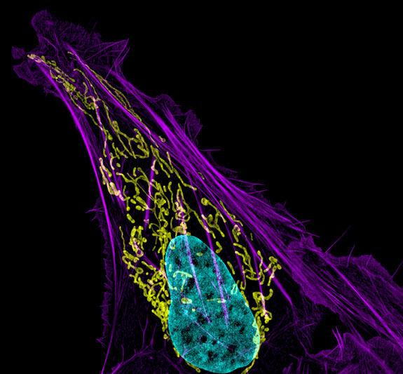
Dylan Burnette, of the National Institutes of Health in Bethesda, Maryland, created this photomicrograph using a technique called structured illumination microscopy (SIM). The image is of human bone cancer (osteosarcoma) showing actin filaments (purple), mitochondria (yellow) and DNA (blue).
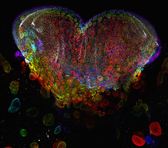
With a confocal microscope, Michael John Bridge, at the HSC Core Research Facilities’ Cell Imaging Lab at the University of Utah, created this close-up of the eye organ of a Drosophila melanogaster (fruit fly) third-instar larvae.
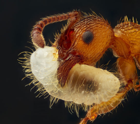
Geir Drange, of Asker, Norway, entered this image of Myrmica sp. (ant) carrying its larva.
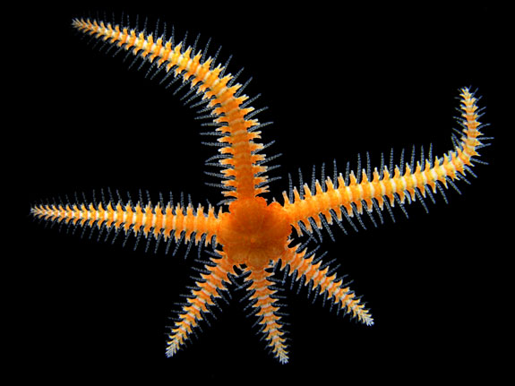
Alvaro Migotto, of the Centro de Biologia Marinha at the University of São Paulo in Brazil, used a combination of stereomicroscopy and darkfield microscopy to capture this brittle star.
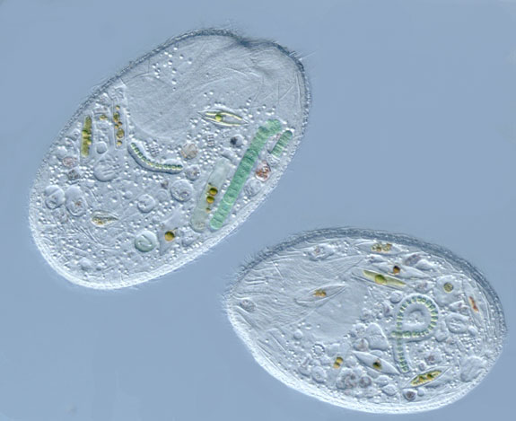
This photomicrograph, by Diana Lipscomb in George Washington University’s Department of Biological Sciences, shows Sonderia sp., a ciliate that preys upon various algae, diatoms and cyanobacteria.
Here, José R. Almodóvar Rivera, of the biology department at University of Puerto Rico’s Mayaguez campus, has captured the pistil, or female reproductive part, of Adenium obesum, a flowering plant native to Africa.
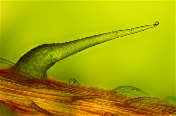
Charles Krebs, of Issaquah, Washington, is a prolific photomicrographer, who has placed in several of Nikon’s competitions. In 2005, he took first prize with an incredible close-up of a house fly. Seen here is a stinging nettle trichome on a leaf vein.
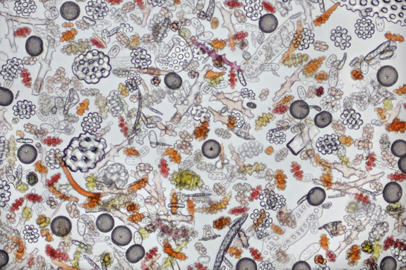
This busy image shows coral sand magnified by 100 times. David Maitland, of Feltwell, England, created it using brightfield imaging.
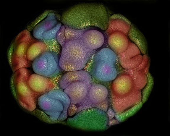
Somayeh Naghiloo, a faculty member of the plant biology department at the University of Tabriz in Iran, submitted this image of the floral primordia of Allium sativum (garlic).
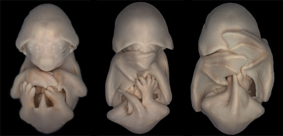
This strangely adorable image of embryos of the species Molossus rufus (black mastiff bat) was taken by Dorit Hockman, of the University of Cambridge’s department of physiology, development and neuroscience.
/https://tf-cmsv2-smithsonianmag-media.s3.amazonaws.com/accounts/headshot/megan.png)


/https://tf-cmsv2-smithsonianmag-media.s3.amazonaws.com/accounts/headshot/megan.png)