The Startling Beauty of the Microscopic
Olympus BioScapes announces ten winners of their 2013 Digital Image Competition, which honors some of the best images taken through a microscope
/https://tf-cmsv2-smithsonianmag-media.s3.amazonaws.com/accounts/headshot/natasha-geiling-240.jpg)
/https://tf-cmsv2-smithsonianmag-media.s3.amazonaws.com/filer/24/3a/243a38f9-51d9-43a6-b729-0ba1568cf741/spike-walker-from-staffordshire-england-won-fourth-prize-for-this-image-it-shows-a-stained-cross-section-of-a-lily-flower-bud.jpg)
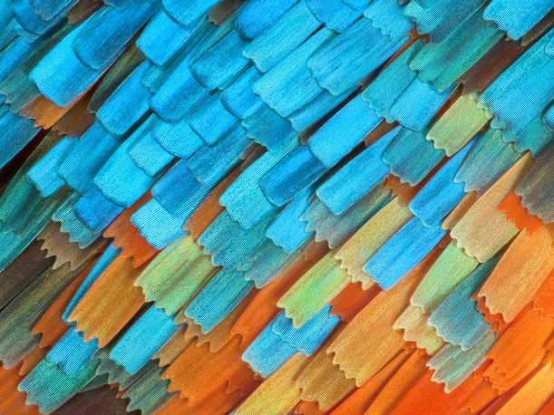
Great purple hairstreak butterfly (Atlides halesus) body scales. Reflected light, 20x. David Millard, Austin, TX, USA. Honorable Mention, 2013 Olympus BioScapes Digital Imaging Competition®.
When Robert Hooke first looked at a piece of cork under a microscope in 1665, he was looking for scientific reasons--but that didn't keep him from seeing the intrinsic beauty in the slides. Of a microscopic flea, he wrote that it was "adorn'd with a curiously polish'd suite of sable Armour, neatly jointed. . ." a description imbued with as much artistic flourish as scientific information.
Since Hooke's time, the connection between microscopic images and artistic value has become thinner--it's easier to think of microscopes as devices of empirical evidence than instruments of artistic inspiration. But occasionally, images come along that combine science and art in such a way to make one wonder whether or not more scientists should approach the study of their microscopic subjects with the same kind of aesthetic eye as Hooke.
Today Olympus BioScapes announced the winners of their 2013 International Digital Imaging Competition, which celebrates some of the world's most amazing pictures taken through the lens of a microscope. "What’s cooler than the intersection of awesome art and serious science?" its website asks, explaining that the goal of the competition is to bring attention to the beautiful stories being told under the microscopes of amateur and professional scientists around the world.
This marks the competition's 10th year, and contestants from 71 different countries submitted over 2100 images and videos. Images could be submitted from any magnification and brand of compound light microscope. From the submitted images, a panel of four individuals (pdf) in the field of microscopy and imaging chose ten images for top prizes, with several dozen others receiving honorable mentions. This year's first place winner, who was awarded a prize of $5,000, went to Dr. Igor Siwanowicz for his photo of a bladderwort, a floating plant that digests microinvertebrates after they touch trigger hairs. Siwanowicz's winning photograph, as well as the nine other top photographers, are shown below.

Humped bladderwort. Confocal imaging, 100x. Igor Siwanowicz, HHMI Janelia Farm Research Campus, Ashburn, VA, USA. First Prize, 2013 Olympus BioScapes Digital Imaging Competition®.
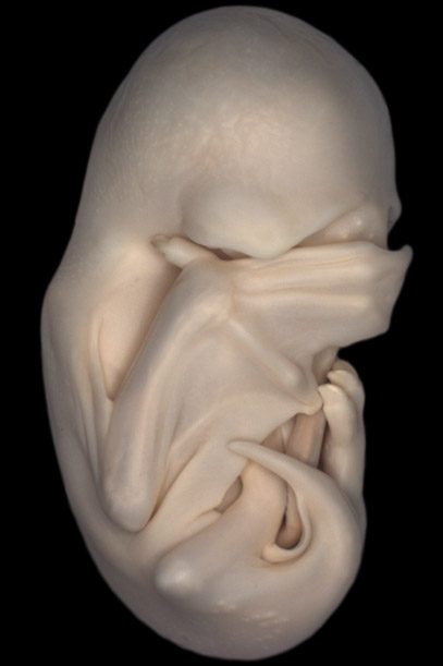
A lateral view of a black mastiff bat embryo (Molossus rufus), at the "Peek-a-boo" stage when its wings have grown to cover its eyes. As development progresses, their fingers grow longer and form the maneuverable struts of their wings, supporting the membrane between their fingers. Stereo microscopy. Dorit Hockman, University of Oxford, Oxfordshire, UK. Second Prize, 2013 Olympus BioScapes Digital Imaging Competition®.
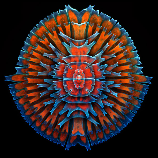
A composite image showing a collection of single-cell fresh water algae, desmids. The red in the image comes from the innate fluorescence of chlorophyll. Confocal imaging, 400x. Igor Siwanowicz, HHMI Janelia Farm Research Campus, Ashburn, VA, USA. Third Prize, 2013 Olympus BioScapes Digital Imaging Competition®.

Mouse embryonic fibroblasts showing the actin filaments (red) and DNA (blue). The image also shows the insides of mitochondria, which were visualized by expressing a green fluorescent protein (GFP) fused to a mitochondrial localization sequence. Structured illumination microscopy (SIM) fluorescence; image acquired with a 60x objective. Dylan Burnette, National Institutes of Health, Bethesda, MD, USA. Fifth Prize, 2013 Olympus BioScapes Digital Imaging Competition®.

"Brother bugs." Gonocerus acuteangulatus, two hours old. 3mm in size. Kurt Wirz, Basel, Switzerland. Sixth Prize, 2013 Olympus BioScapes Digital Imaging Competition®.

Phantom midge larva (Chaoborus) "Glassworm." Birefringent musculature that is usually clear and colorless is made visible here by specialized illumination. Polarized light, 100X. Charles Krebs, Issaquah,WA, USA. Seventh Prize, 2013 Olympus BioScapes Digital Imaging Competition®.

Mouse tail whole mounts stained for the K15 (green) hair follicle stem cell marker as well as Ki67 (red), which marks proliferating cells. Nuclei are marked with DAPI (blue).Technician on the project was Samara Brown. Confocal Z-stack image. Yaron Fuchs, Howard Hughes Medical Institute/The Rockefeller University, New York, NY USA. Eighth Prize, 2013 Olympus BioScapes Digital Imaging Competition®.

Paramecium, showing contractile vacuole and ciliary motion. Paramecium lives in fresh water. The excess water it takes in via osmosis is collected into two contractile vacuoles, one at each end, which swell and expel water through an opening in the cell membrane.The sweeping motion of the hair-like cilia helps the single-celled organism move. Differential interference contrast, 350x-1000x. Ralph Grimm, Jimboomba Queensland, Australia.Tenth Prize, 2013 Olympus BioScapes Digital Imaging Competition®.
/https://tf-cmsv2-smithsonianmag-media.s3.amazonaws.com/accounts/headshot/natasha-geiling-240.jpg)